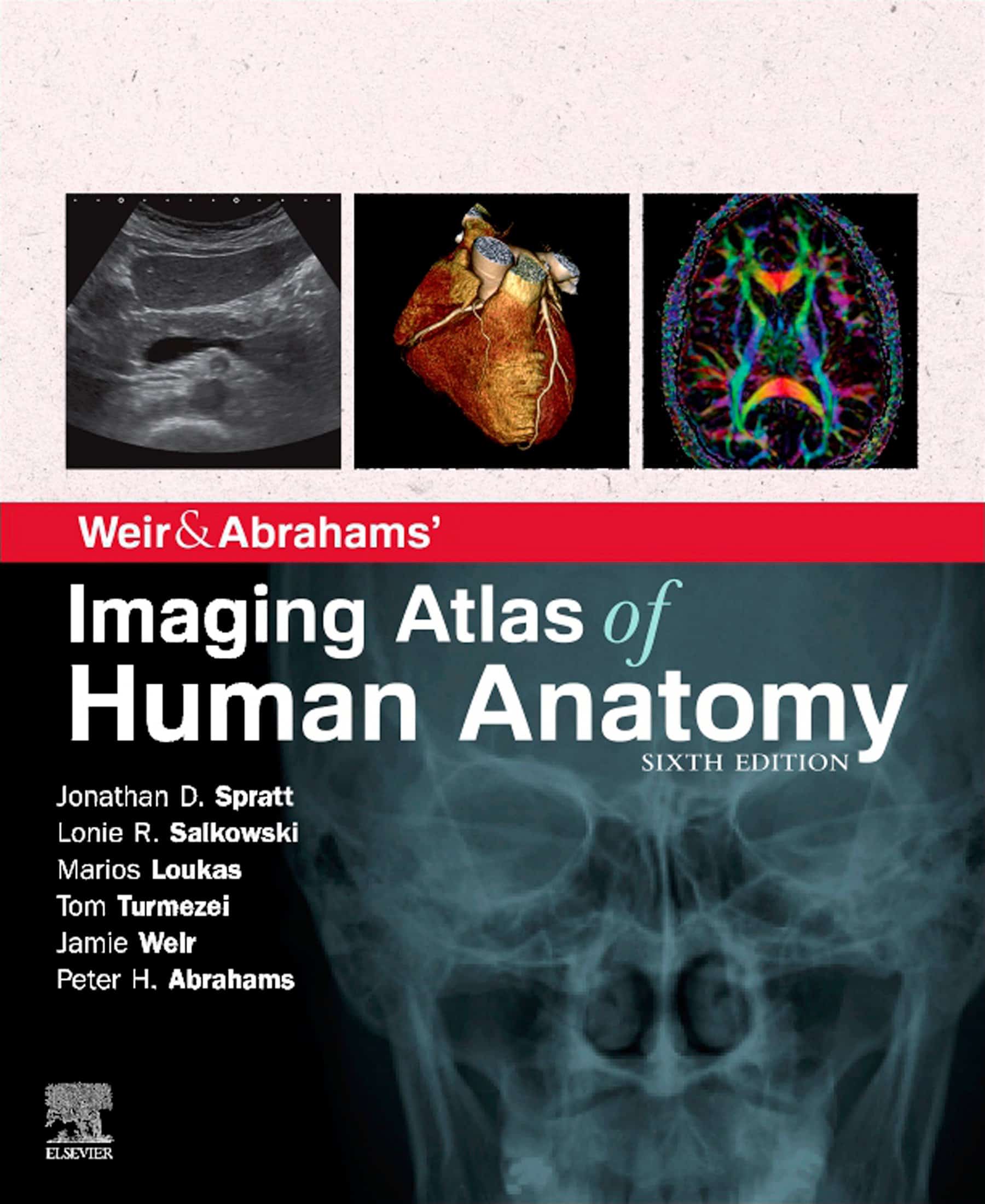
Imaging Atlas of Human Anatomy, 6th Edition, (PDF) is the ideal up-to-date imaging guide for a comprehensive and 3-dimensional understanding of applied human anatomy
Imaging is ever more essential to anatomy education and throughout modern medicine. Building on the success of prior editions, this completely revised sixth edition provides an outstanding foundation for understanding applied human anatomy, providing a complete view of the structures and relationships within the whole body, using very modern imaging techniques.
All related imaging modalities are included, from plain radiographs to more advanced imaging of ultrasound, CT, functional imaging, MRI, and angiography. Coverage is further improved by a carefully selected range of BONUS electronic content, including clinical cases and photos, ultrasound videos, cross-sectional imaging stacks, labeled radiograph ‘slidelines’ and test-yourself materials. Uniquely, key syllabus image sets are now underlined throughout to aid efficient study, in addition to the most common, clinically important anatomical variants that you should be mindful of.
This outstanding package is ideally suited to the requirements of medical students, as well as radiographers, radiologists, and surgeons in training. It will also prove precious to the range of other students and professionals who need an accurate, clear view of anatomy in current practice.
- Completely revised legends and labels and new high-quality images–including the latest imaging techniques and modalities as seen in clinical practice
- Core syllabus image sets now highlighted throughout–to assist you to focus on the most important areas to succeed on your course and in examinations
- New orientation drawings–to help you comprehend the different views and the 3D anatomy of 2D images, in addition to the conventions between cross-sectional modalities
- Exclusive summaries of the most common, clinically important anatomical variants for each body region–shows the fact that around 20% of human bodies have a minimum of one clinically significant variant
- Includes the full variety of relevant modern imaging–featuring cross-sectional views in MRI and CT, ultrasound, fetal anatomy, angiography, plain film anatomy, nuclear medicine imaging, and more – with a better resolution to make sure the clearest anatomical views
- Perfect as a stand-alone resource or in union with Abrahams’ and McMinn’s Clinical Atlas of Human Anatomy–where new links assist put imaging in the context of the dissection room
- Now a complete learning package than ever before, with excellent BONUS electronic enhancements set in within the accompanying eBook, including:
- High-yield USMLE topics–clinical cases and photos for key topics, linked and highlighted in chapters
- Labeled ultrasound videos–bring images to life, showing this increasingly clinically practiced technique
- Questions and answers supplement each chapter–to test your understanding and aid exam preparation
- Labeled image ‘stacks’–that enable you to review cross-sectional imaging as if using an imaging workstation
- Self-test image ‘slideshows’ with multi-tier labeling–to help to learn and cater for beginner to more advanced experience levels
- Labeled image ‘slidelines’–showing characteristics in a full range of body radiographs to enhance understanding of anatomy in this essential modality
- 34 pathology tutorials–based near nine key concepts and illustrated with several additional pathology images, to further build your memory of anatomical structures and lead you through the important relationships between normal and abnormal anatomy
NOTE: The product only includes the ebook Imaging Atlas of Human Anatomy, 6th Edition in PDF. No access codes are included.


Reviews
There are no reviews yet.